The purpose of this research project is to develop a medical imaging system based on microwave imaging suitable for breast-cancer screening. The project covers all aspects of the system including signal processing and hardware manufacturing.
Background
Each year more than 4000 women are diagnosed with breast cancer in Denmark. A key issue in treatment of breast cancer is early detection of the cancer, and the best way to detect the cancer at an early stage is through screening programs. Recent studies of the Danish screening program in Copenhagen have shown a decrease in the mortality rate of as much as 37% for women participating in the screening program compared to those not participating. For a screening program to be effective, the cost of the individual screening must be low and the screening method must have high, operator-independent sensitivity and specificity, and the screening process should be as fast as possible. The term sensitivity is used to describe how many of the potentially malignant tumors the system can detect while specificity is used for describing the ability of the system to distinguish between malignant and the benign tumors (such as cysts). The operator independency is important as nurses and radiographers should be able to perform the examination and hand over the results to a doctor for further analysis. Finally, the speed is important since the screening program should cause as little inconvenience to the patients as possible.
| |
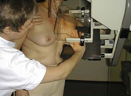
Mammography at Rigshospitalet
|
| |
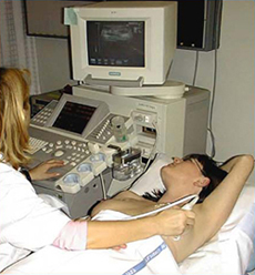
Ultrasound examination at Rigshospitalet
|
At the present, the only suitable screening tool for the detection of breast cancer is X-ray imaging of the breasts, known as X-ray mammography. When using mammography, the breast is illuminated by X-rays and onto a photo-sensitive material. The resulting projection of the X-ray absorption in the breast can then be used for locating tumors. Mammography has proven itself as a very suitable screening tool, especially for postmenopausal women for whom the specificity values higher than 95% have been reported. The sensitivity is, however, often lower (50-80%) and in particular the positive prediction value (i.e., the ratio of the women who are called in for additional examinations who actually have cancer) may be very low. Values as low as 3.3% have been reported.
When mammography screening is combined with a follow-up ultrasonic examination of those women whose mammographies show signs of possible cancer, the sensitivity can reach as much as 95%. Mammography, however, is not suitable for screening of premenopausal women, as the sensitivity for this group of women is as low as 60%. Consequently, the Danish screening program does not include women under the age of 50 years, a group who makes up around 20% of the annual diagnosed breast-cancer cases in Denmark. In addition to the premenopausal women, mammography also performs poorly on the increasing number of postmenopausal women receiving hormonal treatment since this treatment postpone or eliminate many of the physiological changes otherwise associated with the menopause.
To understand the reason for the low sensitivity for younger women it is necessary to realize what is imaged when performing mammography screenings. The mammography is an image of how much of the X-ray radiation is dissipated in the breast. The dissipation of the energy in the X-rays is dependent upon the type of tissue the X-rays need to propagate through from the source to the photo-sensitive detector. Certain types of tissue, such as fat, cause low dissipation while other types, such as tumors and breast tissue, cause high dissipation of the energy of the X-rays. In the breasts of postmenopausal women, the hormonal changes implies that most of the breast tissue has been transformed to fat. Consequently, highly dissipating tissue such as malignant tumors will stand out in the mammographies. For premenopausal women, however, the presence of dense breast tissue means that tumors are more likely to be undetected by X-rays.
As opposed to X-rays, the interaction between microwaves and breast tissue is not governed by the dissipation of energy in the breast tissue, but rather by scattering of the incident microwaves. This scattering is caused by inhomogeneities in the electrical constitutive parameters of the tissue. As early as in the late 1940's it was discovered that the constitutive parameters of malignant breast tissue is 3-10 times that of normal tissue. This difference will cause the microwaves to scatter, and by applying signal processing to the measured scattered waves, the size and position of the inhomogeneities, including malignant tumors, in the breast can be determined. The most intuitive way to present the information extracted from the scattered signal is by producing three-dimensional images. This approach is known as microwave imaging.
Microwave Imaging
The term microwave imaging covers all processes in which measurements of electromagnetic fields in the microwave region from 300 MHz to 30 GHz are used for creating images. To create images from microwave measurements, it is necessary to construct a microwave imaging system, which is able to transmit microwaves and measure the scattered waves at one or more antennas. Different types of microwave imaging systems are currently being used for imaging in such areas as ground penetrating radar and remote sensing. Depending on the items to be imaged, different types of microwave imaging systems are needed. These range from small antennas used for near field measurements in ground penetrating radar to the large airborne systems used in remote sensing. There are two key issues to address when designing a microwave imaging system. One is the increase of the signal to noise ratio in the system and the other is to assure that the system has a large dynamic range. The importance of both of these is closely related to the fact that the scattered signal is often very weak in comparison to the transmitted signal. This implies that any noise in the system will have a large impact on the image quality and that the system must be able to distinguish even small differences in the received signals.
To obtain the maximum amount of information from the microwave measurements, inverse scattering techniques must be applied. The term inverse scattering is used to describe techniques in which the images are created by inverting a model of the scattering mechanisms derived from Maxwell's equations When using inverse scattering for microwave imaging, two things determine the quality of the images. The first thing is the accuracy of the forward model and the other is the accuracy of the inversion algorithm. By using Maxwell equations, an exact solution to the forward scattering problem can be determined. The forward model, however, is often too complex to efficiently be inverted and certain approximations and assumptions must therefore be applied. Consequently, the application of physically viable assumptions and simplifications to the forward model is as important in inverse scattering as the pure mathematical procedure of inverting the model.
Scope of Project
The current research project, which has the objective of creating a prototype imaging system ready for clinical testing, is divided into three major parts:
- Inverse scattering using nonlinear inversion. Due to the complexity of the scattering problem it is necessary to reconstruct the images using full nonlinear inversion. To this end, several different alogrithms have been developed. Special emphasis has been put on the reconstruction speed and robustness of the algorithms.
- Electromagnetic modeling of the imaging system with and without the breast. This is important both to investigate the performance of different setups of the system and because some of the inversion algorithms requires several forward scattering problems to be solved during the image reconstruction.
- Design and construction of the microwave imaging system. Based on the observations made during the electromagnetic modeling of the problem and the results obtained during the development of the inverse scattering algorithms, an imaging system has been constructed. A key factor in the construction of the microwave imaging system has been the reduction of noise in the system.
The TUD Imaging System
The system currently being developed at the Technical University of Denmark is designed for reconstructing images of the interior of the breast using a frequency-domain inverse scattering algorithm. Hence, the system has been designed for operation in the frequency domain from 300 MHz to 3.5 GHz.
When collecting the data an antenna is used to transmit a field of constant frequency while the response is measured by a number of receiving antennas. By comparing the responses measured with an empty system with those obtained when an object is present in the system, the scattered field created by the inserted object is found. This scattered field is then used as the basis for a minimization problem in which the distribution of constitutive parameters which minimizes the difference between a calculated and the measured scattered field is sought for. The minimization problem is formulated using the log-phase formulation and is solved by application of an iterative Newton-based algorithm.
System Setup
The system consists of 32 antennas positioned in a cylindrical setup inside of a measurement tank. During measurements the cylindrical tank is filled with a coupling liquid based on a mixture of glycerin and water. This liquid mimics the electromagnetic parameters of the breast, thereby maximizing the microwave effect coupled to the interior of the breast which, in turn, makes the inverse problem more well behaved. Although the nonlinear inversion problem benefits from the use of the glycerin-water coupling liquid, the lossy implies that the microwave hardware must be able to deal with large differences in the amplitudes of the signals to be measured.
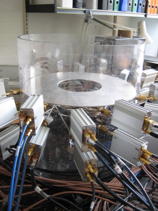
Side view of the prototype microwave imaging system. In the final system, the top of the measurement tank will be cut off and the patient will lie with her breast suspended through the aperture.
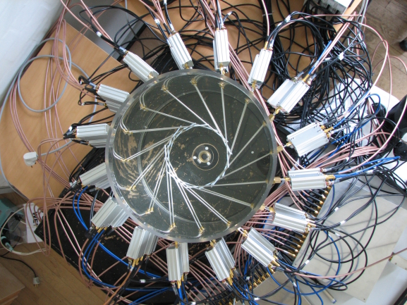
Top view of the prototype imaging system. The horizontal configuration improves the capability of the system to image the region of the breast closest to the chest wall.
Antennas
| |

Photo of one of the antennas of the system. The simple monopole antenna is compact, easy to manufacture, and computational inexpensive to include in electromagnetic models.
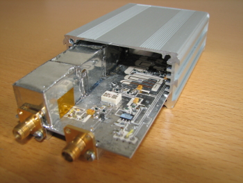
Opened transceiver module.
|
|
The antennas used in the system are of a simple monopole type which is constructed by stripping the outer conductor from the outermost part of a coaxial cable. Such antennas perform poorly when operating in free space but when operating in the lossy coupling liquid, the characteristics of the antenna changes dramatically. In the current configuration with 3.5 cm of the outer conductor stripped from the coaxial cable, the antennas have reflection coefficients of less than -6 dB over the entire frequency band used in the system.
Microwave Hardware
Each antenna is connected to a transceiver module encased in an aluminum box. The transceiver modules are fed by two microwave signals; one feeding the antennas and one, offset by 1 KHz from the feed signal, driving a mixer. The transceiver modules are constructed with special emphasis put on the large dynamic range required by the system. A dynamic control of the amplitude of the signal feeding the antennas which changes the signal level depending on which antenna is receiving further increases the performance of the system.
Milestones
Next Milestone
- January 2009: End of prototype tests. System ready for implementation in exam bed for clinical tests.
Previous Milestones
- September 2008: Preliminary tests end - the imaging system has succeeded in imaging simple objects. Tests of the system in more realistic imaging situations are initiated.
- May 2008: The current prototype is tested for the first time.
- January 2008: An antenna system using horizontally oriented monopoles is build.
- December 2007: A contrast source inversion algorithm is tested for use with the system.
- June 2007: Design of transceiver module is completed.
- April 2007: A imaging system using 32 vertically oriented monopoles is completed.
- December 2006: The Newton-based nonlinear 3-D imaging algorithm is tested for the first time.
- December 2005: The first prototype, based on linear imaging, is discarded due to its incapability to correctly reconstruct the complex scattering environment encountered in breast imaging.
Contact Information
Personnel
-
PostDoc
Tonny Rubæk:Overall system and antenna design, imaging algorithms, and project coordination
-
Also on the project:
Previously Attached Personnel
Funding
Currently, the project is funded by the Villum Kann Rasmussen Foundation, grant no. VKR020891. Additional funding has been received from
- Danish Research Council for Production and Technology Sciences.
- The Otto Mønsted Foundation.
- Valdemar Selmer Trane og Hustru, Elisa Tranes Fond.
- Ulla and Mogens Folmer Andersen's Foundation.
For Students
Students interested in carrying out projects within the scope of this research project should contact Tonny Rubæk for more information. A number of project suggestions can be found in the DTU Project Database.
Previous Special, Midterm, Bachelor, and Master Projects
- Rune Skipper Madsen and Søren Schmidt, "Construction of microwave camera for breast cancer detection", Master project (February 2005 - October 2005).
- Claus Sabroe Nielsen, "Single Arm Archimedian Spiral Antenna for Breast Cancer Detection", Master project (February 2006 - September 2006).
- Andrea Sánchez Amores, "Design of a System of Antennas for Breast Cancer Detection", Master project (September 2006 - April 2007).
Want to Know More ?
On microwave imaging and its applications:
On cancer:
[Back to Research projects]